


- ABSTRACT:
- 1. INTRODUCTION
- 2. METHODS
- 3. RESULTS
- 3.1. Study Profile and Evidence Structure
- 3.2. Thematic Analysis
- 3.2.1. mtDNA Sequence Variants in Recurrent Pregnancy Loss
- 3.2.2. mtDNA Copy Number and Deletion Burden in Pregnancy Loss
- 3.2.3. Endometrial mtDNA Copy Number in Recurrent Implantation Failure
- 3.2.4. Heteroplasmy and Mitochondrial Replacement-Related Evidence in Implantation Failure
- 3.2.5. Embryonic mtDNA Copy Number and Implantation Potential: Positive Evidence
- 3.2.6. Embryonic mtDNA Copy Number and Implantation Potential: Negative and Conflicting Evidence
- 3.2.7. mtDNA Copy Number in Euploid Embryo Transfer, Miscarriage, and Live Birth
- 3.2.8. Embryo Developmental Stage and Sample Source as Modifiers of mtDNA Association
- 3.2.9. Integrated Comparative Synthesis
- 4. DISCUSSION
- STRENGTHS AND LIMITATIONS
- IMPLICATIONS AND RECOMMENDATIONS
- CONCLUSION
- LIST OF ABBREVIATIONS
- AUTHOR'S CONTRIBUTION
- CONSENT FOR PUBLICATION
- AVAILABILITY OF DATA AND MATERIALS
- FUNDING
- CONFLICT OF INTEREST
- ACKNOWLEDGEMENTS
- DECLARATION OF AI
- SUPPLEMENTARY MATERIAL
- APPENDIX
- REFERENCES
Article ID: PD2601205006
Views: 30Mitochondrial DNA Copy Number and Genomic Variants in Recurrent Pregnancy Loss and Recurrent Implantation Failure: A Systematic Review
 PDF
⬇ Downloads: 1
1Patni Charitable Hospital, Karachi, Pakistan
Revised: 05 October, 2026
ABSTRACT:
Introduction: Recurrent pregnancy loss and recurrent implantation failure remain unexplained in a substantial proportion of patients after evaluation for established causes. Mitochondrial DNA number and genomic alterations have been proposed as markers of oocyte competence, embryo development, endometrial receptivity and placental function.
Methods: To synthesize evidence on associations between mtDNA copy number, deletion burden, sequence variants, haplogroups, heteroplasmy, recurrent pregnancy loss, recurrent implantation failure and related reproductive outcomes. Moreover, to distinguish direct evidence from contextual evidence derived from in vitro fertilisation and embryo-transfer populations, PubMed/MEDLINE, Scopus, and Web of Science Core Collection were searched on June 30, 2026.
Results: A total of 29 studies involving primary human studies evaluating mtDNA markers in recurrent pregnancy loss, recurrent miscarriage, early pregnancy loss, recurrent implantation failure or relevant implantation, miscarriage and live-birth outcomes were included. Due to substantial clinical and methodological heterogeneity, we synthesized findings narratively by mtDNA marker type, biological source, and clinical directness. Sequence variants showed the most consistent direct signal among the available direct recurrent pregnancy loss studies.
Conclusion: The available evidence suggests tissue- and context-specific biological and epidemiological associations between alterations in mitochondrial DNA and reproductive outcomes but does not validate mtDNA alterations as diagnostic biomarkers for recurrent pregnancy loss, recurrent implantation failure, or independent embryo-selection tests. Before clinical implementation, prospective multicentre studies using standardized assays, harmonized definitions, proper confounder control, and independent validation are required.
Keywords: Mitochondrial DNA, copy number, recurrent pregnancy loss, recurrent implantation failure, heteroplasmy, embryo implantation, miscarriage.
1. INTRODUCTION
Recurrent Pregnancy Loss (RPL) and Recurrent Implantation Failure (RIF) are different conditions of reproductive failure, which sometimes overlap, but cannot be used interchangeably. Infertility occurs in about one in every six adults around the world [1, 2], whereas recurrent pregnancy loss has severe physical, psychological, and economic implications [3]. These burdens support the clinical interest in exploring other mechanisms in patients whose recurrent reproductive failures are not explained by standard evaluation.
Definitions vary across professional guidelines. ESHRE defines RPL as the loss of two or more pregnancies. In contrast, the RCOG guideline distinguishes between RPL and early miscarriage by retaining the traditional definition of recurrent miscarriage: three or more first-trimester pregnancy losses, but moving the line of investigation of these early miscarriages to earlier in the pregnancy when clinically indicated [4, 5]. No single universally accepted numerical threshold defines Recurrent Implantation Failure (RIF), and its interpretation is influenced by maternal age, embryo competence, the number and quality of embryo transfers, and, where available, embryo ploidy [6].
Established contributors to RPL include chromosomal, uterine, endocrine, immunological, thrombophilic and lifestyle factors, yet a clinically important proportion of cases remains unexplained after standard evaluation [4, 5, 7]. Mitochondrial biology is relevant to this residual group because oocytes, early embryos, endometrium, and placental tissues have stage-specific energy demands and depend on tightly regulated mitochondrial function.
Mitochondria support oocyte competence, fertilisation, early embryonic development, endometrial function and placentation [8–10]. Mitochondrial DNA (mtDNA) copy number reflects the number of mitochondrial genomes in a sample and may vary with mitochondrial biogenesis, cell composition, stress-related compensatory replication, and the normalization strategy used. In contrast, mtDNA sequence variants reflect genomic integrity and may have tissue- and heteroplasmy-dependent effects on oxidative phosphorylation [11–14].
Mitochondrial dysfunction can lead to reproductive failure through a biologically plausible pathway-namely oxidative stress. Mitochondria both generate and respond to Reactive Oxygen Species (ROS), and dysregulated oxidative stress has been associated with miscarriage and other pregnancy complications [15–17]. None of the above is specific to oxidative stress, but mitochondrial dysfunction can also be evaluated using mitochondrial DNA copy number and genetic changes.
Variations in study populations, biological tissues, reproductive outcomes, and assay methods make interpretation of the literature challenging. Few direct RPL/RIF studies exist. However, several larger in vitro fertilisation (IVF) studies compare embryo or trophectoderm mtDNA with implantation or live birth across populations that may not have RPL or RIF. Previous reviews focused on mitochondrial dysfunction or reproductive ageing. Accordingly, this review distinguishes direct RPL/RIF evidence from contextual embryo-transfer evidence and compares quantitative and qualitative changes in mtDNA across biological tissue compartments.
Population-Exposure-Outcome (PEO) framework was used because the review evaluated biomarker associations rather than therapeutic interventions [18]. The population comprised women or couples with RPL, RM, EPL, RIF, or a history of repeated failed embryo transfer. The exposures included quantitative and qualitative mtDNA alterations, including copy number, deletion burden, D-loop variants, coding-region variants, mt-tRNA variants, haplogroups, and heteroplasmy.
Accordingly, the review addressed the following question: Among women or couples with recurrent pregnancy loss or recurrent implantation failure, how are mtDNA copy number and mitochondrial genomic alterations associated with embryo competence, implantation, miscarriage recurrence, and live birth?
The synthesis aimed to investigate mtDNA quantity and genomic integrity; assess the effect of tissue source; and evaluate the clinical relevance of direct evidence over contextual evidence (rather than treating every reproductive study as equally applicable to RPL/RIF).
This review adds value by integrating these strands of evidence within a single tissue- and context-specific framework. Rather than treating mtDNA copy number, sequence variation and embryo-transfer findings as interchangeable evidence, the review distinguishes direct RPL/RIF associations from contextual IVF evidence. It evaluates biological association, prediction, and clinical utility separately.
2. METHODS
2.1. Design and Reporting Framework
This systematic review followed the PRISMA 2020 [19] reporting items. The review was not prospectively registered in PROSPERO; therefore, no registration number is available, and we acknowledge the absence of prospective protocol registration as a methodological limitation. Since the review evaluated associations between mitochondrial genomic markers and reproductive outcomes rather than therapeutic interventions, we planned a structured narrative synthesis before the final synthesis. Meta-analysis was not performed because studies differed substantially in population definitions, tissue source, mtDNA assays, normalization approaches, embryo ploidy control and outcome definitions. Additional limitations include single-reviewer screening and data extraction, heterogeneity in mtDNA assays and biological sources, limited replication of direct RPL/RIF studies, and inability to establish clinical predictive validity.
2.2. PEO Framework and Research Question
The PEO framework comprised of the following elements:
- Population- women or couples with RPL, RM, EPL, RIF, or repeated failed embryo transfer.
- Exposure- mtDNA copy number, deletion burden, sequence variants, haplogroups, or heteroplasmy.
- Outcomes- implantation, embryo competence, miscarriage or recurrent pregnancy loss, clinical pregnancy and live birth.
Studies providing direct RPL or RIF evidence constituted as primary evidence base. Those studies that retained general IVF or embryo-transfer as contextual evidence when they evaluated clinically relevant implantation, miscarriage, or live-birth outcomes. These contextual studies were not used to infer RPL/RIF-specific associations or causality, but to assess whether mtDNA-related biological or predictive patterns observed in embryo-transfer settings were consistent with the direct evidence.
2.3. Eligibility Criteria
Primary human studies in English between 2000 and 2026 that measured an of the mtDNA marker such as copy number or content, deletion burden, sequence variant or haplogroup, or heteroplasmy and correlated it with RPL, RM, EPL, RIF, or a predefined reproductive outcome were qualified in the study. Eligible designs included case-control, cohort, analytical cross-sectional, molecular, and non-randomized intervention or follow-up studies. Reviews, editorials, commentaries, conference-only reports, animal studies, mechanistic studies without a relevant reproductive outcome, and duplicate or overlapping reports that did not provide additional information were excluded. General IVF studies were not counted as specific evidence for RPL/RIF if the study population did not expressly refer to RPL/RIF.
2.4. Information Sources and Search Strategy
PubMed/MEDLINE, Scopus and Web of Science Core Collection were searched using a combination of terms related to mtDNA, copy number/content, variants/mutations, deletion, heteroplasmy, haplogroup, D-loop, mt-tRNA and reproductive failure (e.g. RPL, recurrent miscarriage, RIF, implantation failure, embryo transfer, miscarriage and live birth). The exact strings, fields, and limits for each database are provided in Appendix A and Appendix Fig. (A1–A3).
Searches were limited to human studies published in English between January 2000 and June 2026. Database filters were applied for publication language and document type where available. Only original research articles with extractable primary human data were considered eligible. In addition to database searching, backward citation searching of included studies and relevant reviews was performed by screening reference lists. Forward citation searching was conducted using Scopus and Web of Science Core Collection to identify potentially eligible studies published after the original reports. No additional eligible studies were identified through backward or forward citation searching. All the records retrieved from the citation search were evaluated by the same screening questions. Before duplicate removal, 628 records were identified from the three databases: PubMed/MEDLINE (n = 164), Scopus (n = 333) and Web of Science Core Collection (n = 131). The search screenshots and exact database-specific syntax is specified in Appendix A.
2.5. Study Selection
Title and abstract screening along with full text screening were conducted based on the eligibility criteria. The screening process did not involve duplicate and independent screening, but rather a re-audition of the final set that were included for “publication type” and “relevant” to the eligibility criteria. It is admitted that there is a methodological limitation in the fact that independent duplicate screening is not done. To facilitate the selection process for the final exclusion categories and the number of exclusions, these are detailed in Appendix B and reconfirmed with the PRISMA flow. Appendix Table B1 details the reasons and numbers for exclusion at the title/abstract and full-text stages. The databases-wise identification and duplicate-removal accounting are presented in Appendix Table B2 and the stepwise PRISMA transitions in Appendix Table B3. A citation-level log linking each excluded full-text report to its specific exclusion reason was not retained.
2.6. Data Extraction
Data extraction was performed using the following predefined fields: author/year, country, design, population and sample size, control/comparator, biological material, mtDNA marker, assay method, reproductive outcome, quantitative effect data (where reported) and major limitations/relevance to the review question. Extraction was not independently duplicated. During revision, study-level sample, control, tissue, assay and quantitative outcome information was cross-checked against the available primary full text or authoritative primary article record. Unavailable effect estimates or confidence intervals were recorded as not reported rather than inferred. Supplementary Table S1 provides the study-level verification matrix. The verification process was used to reconcile study-level sample sizes, comparator groups, tissue sources, assays and reported effect estimates before finalising. The PRISMA 2020 study-selection flow is presented in Fig. (1). The database-specific identification is reported in Appendix A, and the merged screening, eligibility and final-study allocation are reported in Appendix B, Table B1–B4. Since the screening and extraction were undertaken by a single reviewer, verification during revision was used to reduce transcription and eligibility errors but does not substitute for independent duplicate reviews.
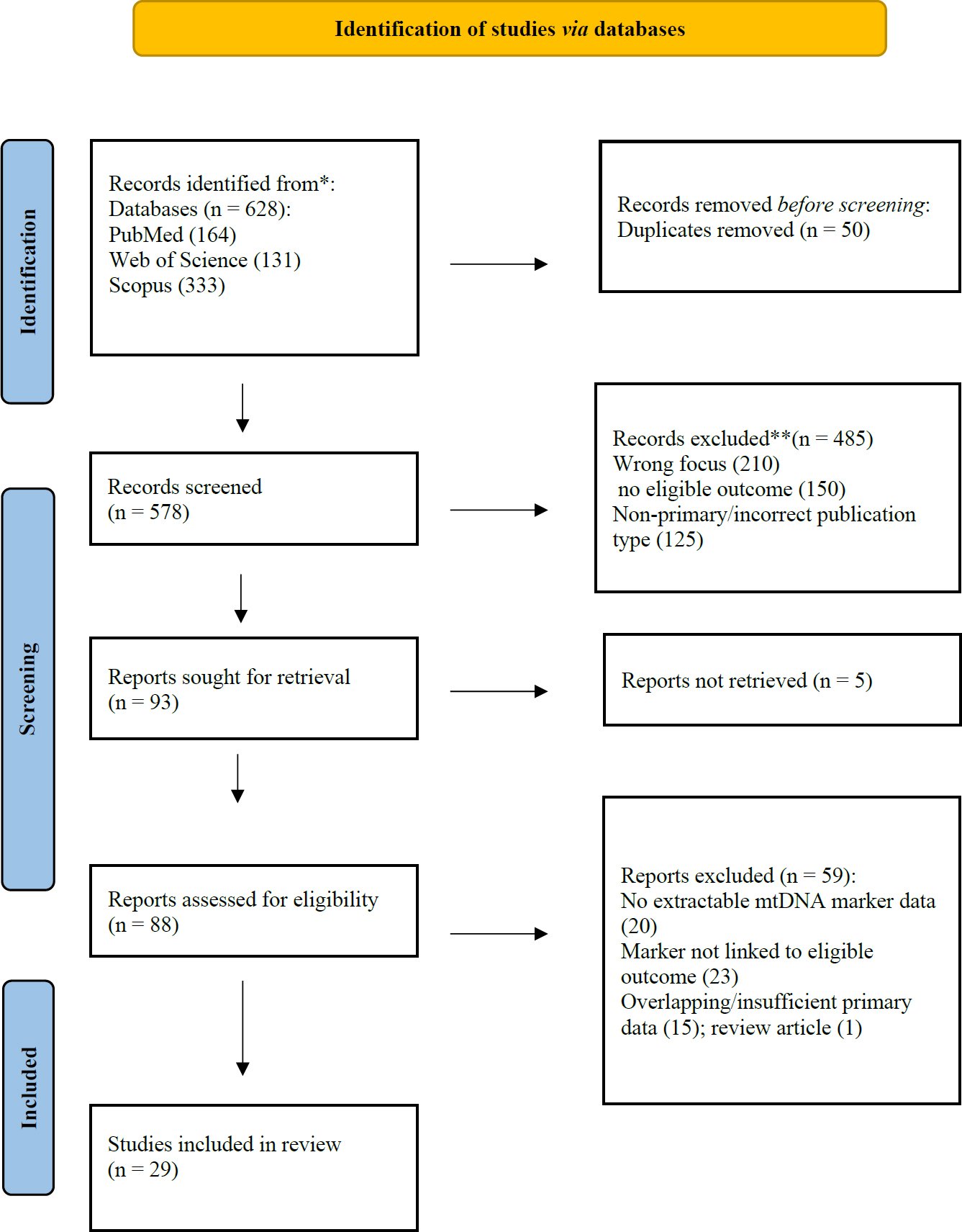
Fig. (1). PRISMA 2020 flow diagram.
2.7. Risk of Bias Assessment
Observational case-control, cohort, analytical cross-sectional, and case-series studies were appraised using the relevant Joanna Briggs Institute (JBI) Critical Appraisal Checklists [20] (Supplementary Table S2A). The two non-randomized ooplasmic-transplantation studies were assessed using ROBINS-I [21] because they evaluated an intervention and follow-up rather than a natural exposure. Domain-level ROBINS-I judgments were presented in Supplementary Table S2B, including confounding, participant selection, intervention classification, deviations from intervention, missing data, outcome measurement, and selective reporting domains.
Risk of bias was evaluated using design-specific tools rather than combining multiple instruments into a composite score (Supplementary Table S2 and S2B). The risk-of-bias appraisal was undertaken and was not independently duplicated. It is acknowledged as a limitation in this study.
The domains under consideration included participant selection, comparability/confounding, validity of exposure or mtDNA measurement, outcome ascertainment, completeness of follow-up where applicable, and appropriateness of statistical analysis. Item-level assessments were completed study by study using the full design-specific JBI checklists (10-item case-control, 10-item case-series, and 11-item cohort checklists) and are reported in Supplementary Table S2A. Only Yes, No, Unclear, and Not applicable response categories were used; no “Partial” category or abbreviated checklist was used. The two non-randomized ooplasmic-transplantation reports were evaluated separately with ROBINS-I and are reported by domain in Supplementary Table S2B. Overall narrative judgements were derived from the pattern and severity of item/domain-level concerns rather than from a numerical score.
Risk-of-bias judgments weight the narrative synthesis, but risk judgments were not used to exclude studies. There was an independent consideration of methodological quality and clinical directness: a study that was more methodologically robust on embryo transfer could give valid evidence on embryo mtDNA but may have been indirect on RPL/RIF as its population was not selected on those grounds. Table 1 presents the design-specific appraisal framework that is employed regarding the included studies. Completed study-specific item-level JBI assessments are presented in Supplementary Table S2A, while domain-level ROBINS-I assessments for the ooplasmic-transplantation reports are presented in Supplementary Table S2B. The JBI appraisal reported confounding factors for some case-control studies and strategies to address confounding, but did not identify the factors. Caution needs to be taken in interpreting these entries, as the latter means that a strategy was reported or judged to be present but does not mean that relevant confounders are fully identified and/or controlled. This apparent discrepancy should not be interpreted as effective confounder control, as it reflects the underlying study reports.
Table 1. Design-specific appraisal framework.
| Study Design | Appraisal Tool | Rationale | Key Domains Assessed |
| Case-control RPL/RM genetic studies | JBI Case-Control Checklist | Direct comparison of affected women with fertile/healthy controls. | Case definition; control selection; comparability; exposure measurement; confounding; analysis. |
| Pregnancy-tissue / early-loss studies | JBI Case-Control Checklist | Cases and control pregnancy tissues were compared for mtDNA markers. | Selection; tissue comparability; assay validity; confounding; outcome classification; analysis. |
| Case series / mutation-screening without controls | JBI Case Series Checklist | Some mutation-screening studies did not include a parallel control group. | Clear inclusion criteria; consecutive/complete inclusion; valid measurement; demographics; reporting. |
| RIF endometrial study | JBI Case-Control Checklist | Endometrial mtDNA was compared between RIF cases and fertile controls. | RIF definition; control appropriateness; cycle timing; assay reliability; confounding. |
| IVF embryo-transfer outcome studies | JBI Cohort Checklist | Embryos/participants were followed from mtDNA measurement to implantation or pregnancy outcome. | Cohort selection; ploidy/embryo selection; exposure measurement; outcome ascertainment; follow-up; confounding. |
| Ooplasmic-transplantation studies | ROBINS-I | Non-randomized intervention/follow-up studies. | Confounding; participant selection; intervention classification; deviations; missing data; outcome measurement; selective reporting. |
Among the 29 primary studies, six were judged at low overall risk of bias (Table 2), 20 at moderate risk and three at high risk. Observational design alone was not treated as an automatic high-risk designation. Recurrent concerns included small or non-disease-specific cohorts, population-specific variant findings without functional replication, retrospective embryo-selection analyses, assay and normalization heterogeneity, incomplete confounder control, and limited external validation.
Table 2. Risk-of-bias summary.
| Overall Risk | Number of Studies | Studies |
| Low | 6 | Fragouli et al. (2017) [22], Ravichandran et al. (2017) [23], Treff et al. (2017) [24], Victor et al. (2017) [25], Lee et al. (2019) [26], Zhou et al. (2021) [27]. |
| Moderate | 20 | Kaare et al. (2009) [28], Seyedhassani et al. (2010a, 2010b) [29, 30], Vanniarajan et al. (2011) [31], Colagar et al. (2013) [32], Azadi et al. (2018) [33], Ye et al. (2020) [34], Ji et al. (2020) [35], Eker et al. (2021) [36], Mojodi et al. (2023) [37], Lledó et al. (2018) [38], Diez-Juan et al. (2015) [39], Fragouli et al. (2015) [40], Stigliani et al. (2014) [41], Klimczak et al. (2018) [42], Łukaszuk et al. (2022) [43], Podolak et al. (2022) [44], El-Damen et al. (2021) [45], Wang et al. (2021) [46], Chuang et al. (2023) [47]. |
| High | 3 | Brenner et al. (2000) [48], Barritt et al. (2001) [49], Victor et al. (2019) [50]. |
Larger embryo-transfer studies generally had stronger outcome ascertainment and, in several cases, controlled embryo ploidy through euploid-transfer designs. Their principal limitation for this review was clinical indirectness because most were not conducted specifically in RPL/RIF populations. Conversely, direct RPL/RIF studies were more clinically relevant but typically smaller and more vulnerable to population stratification, residual confounding, and limited replication.
The risk-of-bias assessment therefore supports cautious, hypothesis-generating interpretation. Neither mtDNA copy-number nor sequence-variant evidence is sufficiently validated for diagnostic or embryo-selection use. Future studies should use prospective multicentre designs, standardized assays, matched controls, prespecified confounder adjustment and independent validation.
2.8. Synthesis Strategy
A meta-analysis was not performed because effect measures and biological substrates were not sufficiently comparable. Narrative synthesis was organized into five prespecified domains:
(1) mtDNA sequence variants in RPL/RM.
(2) mtDNA copy number and deletion burden in pregnancy loss.
(3) Endometrial mtDNA copy number in RIF.
(4) Embryo/trophectoderm mtDNA content and implantation, miscarriage or live birth.
(5) Heteroplasmy and ooplasmic-transfer evidence.
Within each domain, direct RPL/RIF evidence was interpreted separately from contextual IVF evidence. Within each domain, findings were compared for direction and consistency of association, methodological strength, clinical directness and evidence of replication, rather than summarized solely by study-level statistical significance. The PEO framework used to structure the review question is presented in Table 3, and the core concepts used to develop the database searches are summarised in Table 4.
Table 3. Population-exposure-outcome framework used to structure the review question.
| PEO Component | Definition in This Review | Examples |
| Population | Women/couples with RPL, RM, EPL, RIF, or repeated failed embryo transfer | Recurrent miscarriage, recurrent implantation failure, early pregnancy loss |
| Exposure | Quantitative or qualitative mtDNA alteration | Copy number, deletion rate, D-loop variants, tRNA variants, haplogroups, heteroplasmy |
| Outcome | Clinical or embryological reproductive outcome | Embryo quality, implantation failure, miscarriage, live birth |
Table 4. Core search concepts.
| Concept | Example search terms |
| Mitochondrial DNA | “mitochondrial DNA” OR mtDNA OR “mitochondrial genome” OR heteroplasmy OR haplogroup OR “D-loop” OR “copy number” OR deletion |
| Recurrent pregnancy loss | “recurrent pregnancy loss” OR “recurrent miscarriage” OR “repeated pregnancy loss” OR “early pregnancy loss” OR miscarriage |
| Implantation failure | “recurrent implantation failure” OR “implantation failure” OR “failed embryo transfer” OR “embryo transfer outcome” |
| Assisted reproduction outcomes | IVF OR ICSI OR blastocyst OR trophectoderm OR embryo OR “live birth” OR implantation OR miscarriage |
Note: Appendix A provides exact database-specific search strategies for each database.
3. RESULTS
3.1. Study Profile and Evidence Structure
Twenty-nine primary human studies met the criteria for final synthesis. Each included study was assigned to one mutually exclusive evidence category based on its primary population and clinical relevance. The complete study-level allocation is provided in Appendix Table B4. Studies were assigned to mutually exclusive evidence categories based on their primary population and clinical relevance: 10 direct reproductive-failure studies (nine RPL/RM/EPL studies and one direct RIF study), four direct/mixed RIF/RPL intervention or embryo studies, and 15 contextual embryo-transfer, implantation, miscarriage or live-birth studies. A study-to-domain mapping is provided in Appendix Table B4 to ensure transparent allocation without double counting.
A study-level mapping of all 29 included studies is provided in Appendix Table B4. Each study was assigned once according to its primary population and clinical context to avoid double counting. Direct RPL/RM/EPL evidence was prioritised when studies directly investigated recurrent reproductive loss, whereas IVF embryo-transfer studies without a primary RPL/RIF population were classified as contextual evidence.
3.2. Thematic Analysis
3.2.1. mtDNA Sequence Variants in Recurrent Pregnancy Loss
Copy-number alterations may reflect compensatory mitochondrial biogenesis, cellular. composition, developmental stage, or tissue-specific responses rather than a single disease-associated mechanism. Kaare et al. [28] also identified heteroplasmic variants in both cases and controls and found no evidence supporting a strong causal role for maternal heteroplasmic mtDNA mutations. Vanniarajan et al. [31], Colagar et al. [32] and Azadi et al. [33], however, found enrichment of specific mtDNA variants (including Complex-I related variants) in affected populations in Table 5.
Table 5. Characteristics and principal findings of the 29 included primary studies.
| Study / Setting | Population / Sample | mtDNA Marker / Method | Principal Finding | Appraisal and Relevance |
| 1. Kaare et al., 2009 [28] Finland/Europe | Recurrent miscarriage women vs controls; 48 RM + 48 controls | Maternal blood; heteroplasmic mtDNA variants; DHPLC + sequencing | Heteroplasmic mtDNA variants were found in both RM and controls; overall, the study did not support a strong causal role of maternal heteroplasmic mtDNA mutations in recurrent miscarriage. | Strength: Direct RPL topic; good control comparison Limitation: Small sample; mainly maternal blood, not all reproductive tissues Relevance: Use as negative/limited association evidence |
| 2. Seyedhassani et al., 2010 [29] Iran | Repeated pregnancy loss vs controls; 96 RPL + 96 controls | Peripheral blood; mtDNA D-loop mutations; mtDNA deletions; multiplex PCR + sequencing | No mtDNA deletions detected; D-loop point mutations were more frequent in the RPL group. | Strength: Direct RPL + mtDNA control-region analysis Limitation: D-loop only; functional validation absent Relevance: Use in mtDNA D-loop/RPL section |
| 3. Seyedhassani et al., 2010 [30] Iran | 96 women with idiopathic repeated pregnancy loss; no parallel control group in this mutation-screening study | Peripheral blood; mt-tRNA Thr/Pro variants; sequencing | Four mt-tRNA Thr variants were reported: A15907G 2/96 (2.08%), A15924G 3/96 (3.12%), G15928A 10/96 (10.42%), and G15930A 2/96 (2.08%) in the full-text results table; T15972C in mt-tRNA Pro occurred in 1/96 (1.04%). The abstract reports G15930A as 3/96, so we used the full-text table. | Strength: Direct RPL mutation-screening evidence Limitation: No concurrent control group; variants require functional and external validation Relevance: Supportive hypothesis-generating mt-tRNA evidence only |
| Vanniarajan et al., 2011 [31] India | 100 women with RPL + 12 aborted fetal tissues; primary article also reported RPL patients for confirmation and age- and ethnicity-matched fertile controls. | The original report clearly described complete mitochondrial genome sequencing in the discovery cohort comprising 100 RPL women and 12 aborted fetal tissues. The sequencing status of the independent confirmation cohort (55 RPL patients) | Found 681 mtDNA variants, many in ND/Complex-I genes; the study linked these variants with RPL among Indian women. | Limitation: Cohort details and control groups require careful interpretation; the reported control cohort and confirmation groups were considered in the revised appraisal. |
| Colagar et al., 2013 [32] Iran | 33 RPL women + 100 controls | ND1 T4216C mutation; PCR + direct sequencing | T4216C found in 10/33 RPL women = 30% vs 11/100 controls = 11%, p < 0.05. | Strength: Clear case-control comparison with specific mutation Limitation: Small case sample; single variant focus Relevance: Use in ND1/Complex-I mutation section |
| Azadi et al., 2018 [33] Iran | 73 RPL women + 100 controls | Complex-I mtDNA variants; direct sequencing | Reported seven mtDNA complex-I variants including T4216C, A5153G, C10142T, C12062T, A12662G, G14179A, T14263C. | Strength: Direct RPL; variant-focused Limitation: Journal category weaker; functional proof absent Relevance: Use as coding-region mtDNA variant evidence |
| Ye et al., 2020 [34] China | 75 EPL cases + 75 healthy controls | Chorionic villi; mtDNA copy number + 4977-bp deletion rate; qPCR/CNV sequencing | EPL cases had higher mtDNA copy number (p < 0.001) and deletion rate (p = 0.005). Both were independently associated with EPL: CN OR 1.71 (95% CI 1.17-2.49; p = 0.005); DR OR 1.07 (95% CI 1.02-1.12; p = 0.006). | Strength: Direct pregnancy-tissue study with adjusted analysis Limitation: EPL rather than recurrent loss; single-centre cohort Relevance: Core copy-number/deletion evidence for pregnancy loss |
| Ji et al., 2020 [35] China | 150 villous samples: 75 spontaneous abortions + 75 induced abortions | Chorionic villi; mtDNA copy number + heteroplasmy; qPCR + NGS | mtDNA copy number: SA 566 (397–791) vs IA 614 (457–739), p = 0.768; logistic regression: p = 0.196; pathogenic mtDNA mutations higher in SA: 70.7% vs 54.7%, p < 0.05; ND2 mutation: 26.67% vs 13.33%, p = 0.041. | Strength: Strong stats; tissue-specific miscarriage evidence Limitation: Spontaneous abortion, not strictly recurrent loss Relevance: Use as strong miscarriage-tissue mutation evidence |
| Eker et al., 2021 [36] Turkey | 25 women with RIF + 25 fertile controls | Endometrial biopsy; MT-ND1 and MT-CO2 copy number by ddPCR; GAPDH normalization | Normalized MT-ND1 and MT-CO2 copy numbers were higher in RIF endometrium than in fertile controls; Mann-Whitney and ROC analyses were performed. | Strength: Most direct endometrial RIF mtDNA study Limitation: Small single-centre case-control study; no independent threshold validation Relevance: Core but exploratory RIF evidence |
| Mojodi et al., 2023 [37] Iran | 100 women with idiopathic recurrent miscarriage + 100 controls | Peripheral blood; mt-tRNA Cys/Tyr; PCR-SSCP + sequencing | Four variants were identified in cases: 5824C>T (MT-TC), 5836T>C, 5849C>T, and novel 5868T>A (MT-TY); bioinformatic analyses suggested possible pathogenicity. | Strength: Direct recurrent-miscarriage case-control evidence Limitation: Rare variants; population-specific; functional validation and replication absent Relevance: Recent supportive mt-tRNA evidence; not a diagnostic marker |
| Brenner et al., 2000 [48] USA | 23 recurrent implantation failure cases after ooplasmic transfer | Donor-recipient mtDNA heteroplasmy; mtDNA analysis | Demonstrated mtDNA heteroplasmy after human ooplasmic transplantation in RIF context. | Strength: Direct RIF context; clinically important Limitation: Intervention-based; not a natural RIF biomarker Relevance: Use only in RIF heteroplasmy/ooplasmic transfer subsection |
| Barritt et al., 2001 [49] USA | Offspring after ooplasmic transplantation in RIF context; case/follow-up study | Donor/recipient mtDNA; mtDNA fingerprinting | Reported mitochondria/mtDNA inheritance issues in offspring from ooplasmic transplantation. | Strength: Human follow-up; RIF-related heteroplasmy Limitation: Not a conventional RPL/RIF cohort Relevance: Use carefully as heteroplasmy safety evidence |
| Lledó et al., 2018 [38] Spain | 159 non-mosaic euploid embryos from 142 couples undergoing single embryo transfer | Blastocyst/trophectoderm mtDNA analysis; comprehensive mtDNA testing | mtDNA >0.003 was associated with lower ongoing pregnancy (42.96% vs 17.65%, p < 0.05) and higher miscarriage (6.15% vs 40.00%, p < 0.05). Heteroplasmy count was not significantly associated with ongoing pregnancy. | Strength: Euploid single-transfer design with adjusted analysis Limitation: IVF cohort not restricted to RPL/RIF; threshold requires external validation Relevance: Contextual bridge evidence for IVF outcomes |
| Diez-Juan et al., 2015 [39] Spain | 270 single euploid embryo transfers (205 day-3; 65 day-5) plus 10 double-embryo transfers | Embryo mtDNA content; MitoScore/quantification | Higher MitoScore categories showed lower implantation in this cohort. Day-3 embryos with Ms >160 (n = 22) and day-5 embryos with Ms >60 (n = 7) did not implant. | Strength: Euploid-transfer study that generated a testable threshold hypothesis Limitation: Retrospective single-laboratory threshold; later studies did not consistently replicate it Relevance: Historical positive evidence; interpret with later contradictory studies |
| Fragouli et al., 2015 [40] UK/USA | 379 embryos (39 cleavage-stage and 340 blastocysts) | Embryo/trophectoderm mtDNA level; NGS/PGT-related analysis | Altered mtDNA levels associated with female age, aneuploidy, and implantation potential. | Strength: High-quality genetics journal; mechanistic relevance Limitation: Embryo-focused, not RPL/RIF-specific Relevance: Use in implantation potential section |
| Stigliani et al., 2014 [41] Italy | 699 day-3 embryo spent-culture-medium samples | Spent culture medium mtDNA/gDNA ratio; non-invasive quantification | Higher spent-medium mtDNA/gDNA ratio was associated with blastocyst development (p = 0.0251) and implantation of good-quality embryos (p = 0.0452). | Strength: Large non-invasive culture-medium dataset Limitation: Extracellular mtDNA is not equivalent to intracellular embryo mtDNA; contamination/standardization concerns Relevance: Contextual evidence for a distinct non-invasive compartment |
| Fragouli et al., 2017 [22] UK/USA | 199 transferred euploid blastocysts from 174 couples | Trophectoderm mtDNA quantification | 190/199 had mtDNA below the prespecified threshold; 9/57 non-implanting embryos had elevated mtDNA, and none of the elevated group implanted. Ongoing pregnancy was 64% with normal mtDNA vs 0% with elevated mtDNA (p < 0.0011). | Strength: Blinded prospective non-selection design Limitation: Single-clinic transfer context and threshold portability remain concerns Relevance: Strong positive prospective evidence, balanced by later negative studies |
| Ravichandran et al., 2017 [23] USA | 1505 euploid blastocysts from 490 couples, 35 IVF clinics | Trophectoderm mtDNA level; qPCR after PGT-A/WGA | Among 282 transferred euploid blastocysts, 249 had normal mtDNA and 185 implanted (74.3%); none of the 33 blastocysts with elevated mtDNA implanted (p < 0.0011). | Strength: Large multicentre euploid embryo dataset Limitation: Embryo-focused, not direct RIF-only Relevance: Use as strong implantation-failure biomarker evidence |
| Treff et al., 2017 [24] USA | 187 patients; 374 sibling euploid blastocyst biopsies, with paired implantation comparison in the informative sibling-outcome subset | Trophectoderm mtDNA level | Found mtDNA levels did not predict reproductive potential of sibling embryos. | Strength: Strong negative comparator evidence Limitation: Embryo-only, not RPL/RIF cohort Relevance: Use as contradictory evidence |
| Victor et al., 2017 [25] USA/UK | 1,396 blastocysts from 259 patients; qPCR validation in 150 samples | Adjusted mtDNA quantification | After correction for chromosomal/genetic factors, mtDNA levels were broadly uniform and did not meaningfully differ by ploidy, maternal age, or implantation potential. | Strength: Large dataset with NGS and qPCR comparison Limitation: Retrospective single-centre analysis; method differences limit cross-study comparability Relevance: Key negative evidence against a universal mtDNA threshold |
| Klimczak et al., 2018 [42] USA | 259 patients; 1,510 blastocyst biopsies (717 euploid embryos in restricted analysis) | Embryonal mtDNA content | Higher mtDNA was associated with poorer embryo grade in the full dataset (p = 0.003), but the association disappeared in euploid embryos (p = 0.834); maternal age showed negligible correlation. | Strength: Large embryo dataset with euploid subgroup analysis Limitation: Retrospective; embryo-focused rather than RPL/RIF-specific Relevance: Shows confounding by ploidy/embryo characteristics |
| Łukaszuk et al., 2022 [43] Poland | 716 embryos from 275 PGT-A patients; includes RIF ≥2 and recurrent miscarriage ≥2 indications | Trophectoderm mtDNA/nDNA ratio from NGS data | Aneuploid embryos had higher mtDNA than euploid embryos, p = 0.02; day-5 blastocysts higher than day-6, p = 0.001; clinical outcome difference not significant, p = 0.39. | Strength: Includes RIF/RM indications; detailed embryo stats Limitation: Outcome association weak/non-significant Relevance: Use as supportive RIF/RM indication + implantation evidence |
| Podolak et al., 2022 [44] Poland | 69 couples; 314 cleavage-stage embryos | Cleavage-stage embryo mtDNA copy number | mtDNA content differed by chromosomal status (reported p < 0.004) but did not show a robust association with embryo morphology; final reproductive associations were not sufficiently consistent for clinical prediction. | Strength: Cleavage-stage NGS dataset Limitation: Retrospective single-centre study; stage-specific and not RPL/RIF-specific Relevance: Contextual developmental-stage evidence |
| Lee et al., 2019 [26] Taiwan | 1,617 embryos from 380 infertile couples; 267 transferred blastocysts analysed for implantation | Adjusted embryo mtDNA quantity | Implanted (n = 103) and non-implanted (n = 164) blastocysts had similar adjusted mtDNA (median 0.00097 vs 0.00088; p = 0.21). | Strength: Large double-masked prospective observational dataset Limitation: Single Asian centre; mtDNA associated with ploidy but not implantation Relevance: Strong negative evidence for implantation prediction |
| Victor et al., 2019 [50] USA/UK | 109 transferred blastocysts in 100 women | Trophectoderm mtDNA content | mtDNA did not differ significantly between implanted (n = 55) and non-implanted (n = 54) blastocysts; very high mtDNA did not preclude healthy pregnancy and birth. | Strength: Direct transfer-outcome counterexample to exclusion thresholds Limitation: Selected transfer cohort; retrospective Relevance: Important evidence against deselecting embryos solely for high mtDNA |
| El-Damen et al., 2021 [45] UAE/IVF setting | 355 single euploid frozen embryo transfers in 314 patients | Blastocyst/trophectoderm mtDNA content | Pregnancy rate 66.2% (235/355); clinical pregnancy 52.4% (186/355); miscarriage 5.6% (20/355). mtDNA did not differ between pregnant/non-pregnant transfers (p = 0.095) or clinical pregnancy/miscarriage (p = 0.136). | Strength: Single-euploid transfer with miscarriage endpoint Limitation: Retrospective; not RPL-specific Relevance: Strong negative evidence for miscarriage prediction |
| Zhou et al., 2021 [27] China | 316 euploid blastocysts from 233 couples; includes RIF/RM indications | Trophectoderm mtDNA content; NGS PGT-A data | No mtDNA difference across live-birth, miscarriage, and non-implantation groups (p = 0.999); no association with implantation (p = 0.965) or adjusted live birth (p = 0.567). Elevated vs normal mtDNA live-birth rates were 26.7% vs 33.6% (p = 0.780). | Strength: Live-birth endpoint; euploid embryo control Limitation: Not purely RPL/RIF-only Relevance: Use as negative live-birth biomarker evidence |
| Wang et al., 2021 [46] China | 769 euploid blastocysts in primary analysis; day-5 n = 355, day-6 n = 388, day-7 n = 26 | Trophectoderm mtDNA content | Aneuploid blastocysts had higher mtDNA (p < 0.001). Day-5 euploid blastocysts had higher mtDNA than day-6/day-7 (p < 0.001); better trophectoderm quality was associated with lower mtDNA (p = 0.026). Lower mtDNA in day-5 euploid blastocysts was associated with better implantation/live birth, but mtDNA was not supported as an independent predictor. | Strength: Large euploid embryo dataset Limitation: Retrospective; embryo-development effects complicate clinical interpretation Relevance: Contextual positive association with explicit limits on prediction |
| Chuang et al., 2023 [47] Taiwan | 1,675 embryos from 1,305 individuals; 1,340 euploid single-transfer outcomes analysed | Embryo/trophectoderm mtDNA content | Day-6 eSET (n = 139) showed lower mtDNA in adverse outcome groups (fetal heartbeat p = 0.004; ongoing pregnancy p = 0.007; live birth p = 0.01); no corresponding association was observed in day-5 eSET (n = 1,201). | Strength: Large stratified euploid-transfer dataset Limitation: Retrospective; association confined to day-6 subgroup Relevance: Strong evidence that developmental day modifies mtDNA-outcome associations |
The sequencing approach should be interpreted according to the cohort structure reported by Vanniarajan et al. [31]. Complete mtDNA sequencing was described for the primary investigated RPL cohort and fetal tissues, whereas the extent of sequencing within the confirmation and control groups was not clearly specified in the original publication.
The D-loop and mt-tRNA studies were similarly hypothesis-generating and were limited by their population-specific contexts. Seyedhassani et al. [29] reported a higher frequency of D-loop point mutations in women with repeated pregnancy loss, whereas the separate Seyedhassani et al. mt-tRNA Thr/Pro mutation-screening study [30] included 96 women with idiopathic RPL and no concurrent control group. Mojodi et al. [37] determined four mt-tRNA-Cys/Tyr variants in 100 Iranian women with Idiopathic Recurrent Miscarriage (IRM) and 100 controls. These findings do not necessarily imply universally causal mutations; rather, they identify candidate susceptibility loci that require further functional validation and external replication.
3.2.2. mtDNA Copy Number and Deletion Burden in Pregnancy Loss
Level of pregnancy-related copy number and deletions were not even. Ye et al. [34] analysed 75 EPL cases and 75 controls and reported significant increased copy number of mtDNA and increased chorionic-villus deletion rate in the EPL cases; in the logistic regression analysis, the copy number of mtDNA was independently associated with EPL (OR 1.71, 95% CI 1.17-2.49), as was the increased degree of chorionic-villus deletion (OR 1.07, 95% CI 1.02-1.12). In contrast, Ji et al. [35] found no significant difference in copy numbers between villus from spontaneous abortions and villus of induced abortions and potentially pathogenic mutations in the mtDNA were more common in the spontaneous abortion. These two studies pinpoint two different biological processes with the amount of mtDNA and the integrity of mtDNA genome.
The clinical significance of this difference is of interest. The number of copies may be affected by mitochondrial biogenesis, cell composition, trophoblast stress and normalization method; while sequence level changes may offer a more specific reflection of the integrity of the mitochondrial genome. Both should only be used in conjunction with the source of the tissue, embryonic chromosome status, and/or analytic method.
3.2.3. Endometrial mtDNA Copy Number in Recurrent Implantation Failure
Direct evidence of RIF was limited. Eker et al. [36] performed an analysis of 25 women with RIF and 25 fertile women by ddPCR normalised to the GAPDH gene for both MT-ND1 and MT-CO2. The copy-numbers of both mitochondrial targets were elevated in the RIF group. This is a clinically relevant study as the endometrium is directly investigated; however, the small sample size and single-centre case-control study design renders the results preliminary and further validation in larger independent cohorts is necessary.
The level of Endometrial mtDNA copy number may be affected by cycle timing, cell composition, the presence of inflammation and normalization strategy. Eker et al. [36] therefore report some preliminary evidence of an association between mitochondrial state in ECL and the diagnosis of RIF, though the study is small and cross-section at one site, does not so far reproduce a diagnostic threshold, predicts the diagnosis in an independent cohort, or shows clinical usefulness.
3.2.4. Heteroplasmy and Mitochondrial Replacement-Related Evidence in Implantation Failure
Brenner et al. [48] and Barritt et al. [49] provide intervention-based evidence from cytoplasmic transplantation in patients with prior implantation failure. These studies demonstrated donor-recipient mtDNA heteroplasmy after cytoplasmic transfer and in resulting offspring. Their relevance to this review is biological and safety-related; they do not estimate the prevalence or diagnostic significance of endogenous heteroplasmy in natural RIF.
Lledó et al. [38] evaluated 159 non-mosaic euploid embryos from 142 couples undergoing single embryo transfer. Elevated mtDNA above the study threshold was associated with a lower ongoing-pregnancy rate (42.96% vs 17.65%, p < 0.05) and a higher miscarriage rate (6.15% vs 40.00%, p < 0.05), while heteroplasmy burden was not significantly associated with ongoing pregnancy. Because the cohort was not restricted to RPL/RIF, this study is best considered bridge evidence rather than direct RIF evidence.
3.2.5. Embryonic mtDNA Copy Number and Implantation Potential: Positive Evidence
Several early studies reported an inverse association between embryonic mtDNA content and implantation. For example, no embryos with the highest mtDNA levels implanted, and Diez-Juan et al. [39] similarly reported declining implantation rates with increasing MitoScore. Ravichandran et al. [23] similarly reported that none of the 33 transferred euploid blastocysts with mtDNA levels above the study threshold implanted (p < 0.0011). Fragouli et al. [40] also reported associations between mtDNA levels and aneuploidy, implantation potential, and maternal age.
A distinct biological compartment was examined through analysis of spent culture medium. Stigliani et al. [41] analysed 699 day-3 spent-culture-medium samples and found that the mtDNA/gDNA ratio was positively associated with blastocyst formation (p = 0.0251) and implantation of good-quality embryos (p = 0.0452). Findings derived from spent culture medium should not be interpreted as equivalent to measurements of intracellular embryonic mtDNA. Wang et al. [46] reported associations between mtDNA content and embryo characteristics and outcomes among day-5 euploid blastocysts; however, the findings did not support the use of mtDNA as a stand-alone predictor.
3.2.6. Embryonic mtDNA Copy Number and Implantation Potential: Negative and Conflicting Evidence
Subsequent studies challenged the reproducibility of a universal mtDNA threshold. Treff et al. [24] found that trophectoderm mtDNA levels did not predict the reproductive potential of sibling embryos. Victor et al. [25], analysing 1,396 blastocysts from 259 patients, reported broadly uniform mtDNA levels irrespective of ploidy, maternal age or implantation potential. Lee et al. [26] analysed 1,617 embryos from 380 couples and found no significant mtDNA difference between implanted and non-implanted transferred blastocysts (median 0.00097 vs 0.00088; p = 0.21). These negative studies directly weaken the case for using a single mtDNA threshold to discard embryos.
The conflicting embryo literature is plausibly explained by both methodological and biological heterogeneity: qPCR versus NGS-derived ratios; effects of whole-genome amplification; different normalization algorithms; day-3 versus day-5/day-6 sampling; untested, aneuploid or euploid embryos; fresh versus frozen transfer; and inconsistent outcome definitions. Consequently, a threshold derived in one laboratory or embryo-stage context cannot be assumed to transfer to another.
3.2.7. mtDNA Copy Number in Euploid Embryo Transfer, Miscarriage, and Live Birth
Euploid-transfer studies are especially informative because they reduce confounding by embryo aneuploidy. Fragouli et al. [22] prospectively evaluated 199 transferred euploid blastocysts and found that all nine embryos with mtDNA above the prespecified threshold failed to implant; ongoing pregnancy was 64% in the normal-mtDNA group and 0% in the elevated group (p < 0.0011). However, later euploid-transfer studies produced non-significant or directionally different results, demonstrating that ploidy control alone does not resolve assay and cohort heterogeneity.
El-Damen et al. [45] analysed 355 single euploid frozen embryo-transfer cycles and found no significant difference in mtDNA content between pregnant and non-pregnant transfers (p = 0.095) or between clinical pregnancies and miscarriages (p = 0.136); mtDNA therefore did not predict miscarriage. Zhou et al. [27] likewise concluded that mtDNA content was not a reliable live-birth screening biomarker after single euploid blastocyst transfer. Victor et al. [50] reported 109 transferred blastocysts and found no significant mtDNA difference between implanted (n = 55) and non-implanted (n = 54) embryos, including healthy births from embryos with very high mtDNA. These data argue against using mtDNA content as an exclusion criterion.
3.2.8. Embryo Developmental Stage and Sample Source as Modifiers of mtDNA Association
Developmental stage is an important modifier. Podolak et al. [44] studied 314 cleavage-stage embryos from 69 couples and found significant differences in mtDNA content by chromosomal status but no robust relationship with morphology or final clinical outcome. In 716 embryos from 275 PGT-A (Preimplantation Genetic Testing for Aneuploidy) patients, Łukaszuk et al. [43] found higher mtDNA in aneuploid than euploid embryos (p = 0.02) and higher values in day-5 than day-6 blastocysts (p = 0.001), while the clinical-outcome difference was non-significant (p = 0.39). Klimczak et al. [42] similarly found an association between mtDNA and poorer embryo grade across 1,510 biopsies (p = 0.003), but the association disappeared when analysis was restricted to 717 euploid embryos (p = 0.834).
Chuang et al. [47] further demonstrated stage dependence: among single-euploid transfers, lower mtDNA ratios were associated with adverse outcomes in day-6 embryos (fetal heartbeat p = 0.004; ongoing pregnancy p = 0.007; live birth p = 0.01), whereas no corresponding association was observed in the much larger day-5 transfer group. Collectively, these findings suggest that mtDNA content is more closely tied to embryo developmental context than to a universally reproducible clinical endpoint.
3.2.9. Integrated Comparative Synthesis
Across the direct evidence, sequence-variant studies report the most consistent RPL/RM signal, but positive findings are generally small, population-specific, and incompletely replicated. Direct RIF evidence is much more limited and rests mainly on one small endometrial case-control study. Embryo-transfer studies are methodologically larger and often better controlled for ploidy, but they are usually indirect for RPL/RIF.
Accordingly, internal validity and clinical directness were interpreted separately. General IVF findings were used to evaluate biological plausibility and prediction in embryo-transfer settings, not as evidence of RPL/RIF causality.
Across embryo studies, early threshold-based associations were not consistently replicated in sibling-embryo designs, adjusted quantification studies, or later euploid-transfer cohorts. Consequently, the evidence does not support mtDNA copy number as a universal threshold, a stand-alone embryo-selection test, or a diagnostic test for RPL/RIF.
Clinical meaning also differs by level of inference. Accordingly, the synthesis prioritised evidence of reproducibility, discrimination of clinically relevant outcomes and demonstrated improvement in patient management over statistical significance alone. A between-group mtDNA difference represents a biological association; an adjusted statistical relationship with a reproductive outcome represents an epidemiological association; reproducible discrimination of future implantation, miscarriage or live birth represents a predictive association; and evidence that using the marker improves selection, management or patient outcomes would establish clinical utility. The included literature contains examples of biological and epidemiological associations, inconsistent evidence for prediction, and no convincing evidence that mtDNA-guided management improves clinical outcomes. Based on this, studies with small, population-specific findings were interpreted as hypothesis-generating unless there was support either through replication or more powerful comparative designs and were not given greater evidential weight because they were found statistically significant. In order not to mix up the concepts of statistical significance and clinical usefulness, the discussion separates these four levels of evidence in a very reflective manner (Table 6).
Table 6. Clinical interpretation of the synthesis by evidence domain.
| Evidence Domain | Biological Association | Epidemiological Association | Predictive Association | Clinical Utility |
| Sequence variants in RPL/RM | Variant enrichment and recurrent locus-specific findings reported in several direct studies. | Some case-control differences were statistically significant, but replication across populations is limited. | No externally validated model predicts future miscarriage/RPL from these variants. | No evidence that variant-guided testing improves management or live-birth outcome. |
| Pregnancy-tissue mtDNA copy number/deletion | EPL tissue studies show altered copy number/deletion or mutation burden in some cohorts, but findings are not uniform. | Ye et al. [34] reported adjusted associations for copy number and deletion rate; Ji et al. did not find a copy-number association. | No prospective, externally validated prediction of future loss. | No validated diagnostic threshold or management benefit. |
| Endometrial mtDNA copy number in RIF | One small case-control study reported higher MT-ND1/MT-CO2 copy number in RIF endometrium. | The same small sample reported a group-level association, with ROC analysis. | No independent threshold validation or prospective prediction. | Clinical utility is unproven; not ready for diagnostic use. |
| Embryo/trophectoderm mtDNA content | Early studies reported threshold-related differences; later studies showed strong dependence on ploidy, developmental day, and quantification method. | Associations with implantation/live birth are inconsistent after controlling for embryo characteristics. | Threshold performance was not reproducible across sibling-embryo and euploid-transfer cohorts. | No evidence that mtDNA-guided selection improves outcomes over established embryo-selection approaches. |
| Heteroplasmy / ooplasmic-transfer evidence | Human ooplasmic-transfer studies demonstrate donor-recipient heteroplasmy and inheritance of donor mtDNA. | These intervention studies do not establish an endogenous association between RPL/RIF and disease. | No validated prediction of implantation or recurrent loss. | Relevant mainly to biological/safety interpretation, not RPL/RIF diagnostic utility. |
Note: Biological association = a difference in mtDNA biology between groups; epidemiological association = a statistical relationship with an outcome; predictive association = reproducible discrimination of a future outcome; clinical utility = evidence that using the marker improves selection, management, or patient outcomes.
These data support a tissue-specific model rather than a single mitochondrial biomarker. Maternal blood can indicate inherited background differences, pregnancy tissue can indicate loss-related mutation load, the endometrium may indicate receptivity-related mitochondrial strain, and embryo or culture-media measurements can indicate development. Future researchers ought to have a prospective test of these compartments instead of assuming that the compartments are interchangeable.
4. DISCUSSION
The data currently available suggests that association of mtDNA changes with reproductive outcomes is condition and method specific and depends on the type of biological tissue analyzed. Though direct RIF evidence is limited to rather small endometrial datasets, evidence of sequence level variation according to direct RPL was reported more often. Embryo-transfer studies were most convincing, but not consistent and in many studies populations did not meet the definition of RPL/RIF.
Results of direct reproductive-loss studies point to higher uniformity of the alterations of the sequence of mtDNA than of their copy number. The increased copy-number and deletion levels observed in Early Pregnancy Loss (EPL) tissue in Ye et al. [34] were not seen by Ji et al. [35] who found an increased frequency of potentially pathogenic variants but did not observe a significant difference in copy-number of mtDNA between EPL and induced abortion tissues. The conflicting results suggest that the amount of mtDNA and the integrity of the mtDNA genome are two distinct biological signatures and hence should not be considered as synonymous.
To differentiate between what is clinical “useful” and what is statistically significant, these 4 grades of evidence are clearly differentiated (Table 6). In some studies, there were indications of biological or epidemiological differences; however, the predictive value of mtDNA- based evaluation of non-invasive measurements for reproductive outcomes remains to be evaluated and no study has borne out that a diagnosis based on mitochondrial DNA or embryo selection results in higher live birth rates or lower rates of miscarriage or alters care compared to standard care. It must be evident that recorded p-values, odds ratio analysis or ROC analysis cannot prove clinical utility.
There is a tissue-specific pattern since failure of the reproduction process may occur at various points, such as embryo competence before implantation, interaction between the embryo and the endometrium during implantation, and maintenance of pregnancy after implantation. Numerous differences in the requirements of the mitochondria exist among these stages, so that it is not likely that the same measurement of mtDNA would have the same meaning in maternal blood, villous tissue, endometrium, trophectoderm, and spent culture medium.
Of the direct RPL studies, mtDNA sequence variation was more consistent than copy number changes, and more variation existed among copy-number findings based on the biological context, tissue source and analytical techniques. In view of these modest successes by Kaare et al. [28] and the population specific nature of each of these studies, as well as the fact that most of the identified variants have not yet been functionally validated, it seems an inherently weak claim to describe any of these as simple causes of RPL.
A more conservative view is that the variants of mtDNA could affect susceptibility among specific populations. Association is necessary but not sufficient evidence for biological plausibility of complex I and mitochondrial translation variants as functional pathogenicity. Results need to be reproduced in other ancestry groups and have to be confirmed on a functional level.
Additionally, genome integrity and copy number have been shown to be uncoupled in studies of pregnancy tissue. EPL has a higher number of mtDNA copy number and mtDNA deletion burden than control in Ye et al. [34] but not found by Ji et al. [35] and they reported a higher frequency of potentially pathogenic mtDNA variants in spontaneous abortions compared to control. The results of these studies do not necessarily contradict each other since the quantity and mutational load of mtDNA would embody different aspects of mitochondrial biology.
There is limited direct evidence for RIF, which has not yet been translated to the clinic. With fewer individuals being studied (25 cases and 25 controls), Eker et al. [36] reported higher copy counts of endometrial-MT-ND1 and MT-CO2 in RIF. A diagnostic biomarker would need to be independently validated, have prespecified thresholds, and have a consistency of performance between laboratories, in addition to being reproducible and cycle timed, which would also require that endometrium be pathological.
Ooplasmic-transfer follow-up studies show that donor and recipient mitochondrial populations may persist in the oocytes, embryos, or offspring. These studies support the biological plausibility of heteroplasmy and the safety concerns, however they are not indicative of a common or diagnostic characteristics of RIF.
The risk of prematurely translating biomarkers containing risks has been reported in the literature related to embryo transfer. Preliminary studies using threshold criteria identified a high negative predictive value of high mtDNA. Later prospective studies and sibling-embryo studies, adjusted-quantification and euploid-transfer studies, however, have failed to demonstrate such a general relationship. Any changes in assay platform, day of the assay, embryo selection, embryo normalisation or transfer conditions will affect the apparent direction and strength of association.
Embryo transfer studies have not provided consistent evidence of correlation between increased mtDNA content and/or reproductive outcome. An important counter example does exist; according to Victor et al. [50] embryos containing high levels of “mtDNA” may lead to healthy pregnancies and healthy births. Unlike these findings, Lee and colleagues [26] reported no significant correlation between adjusted embryo mtDNA content with implantation, while Zhou and colleagues [27] found no association between embryo mtDNA content with euploid embryo outcomes of implantation or miscarriage or live birth. Overall, these results indicate that embryo mtDNA quantity is not likely to be an independent predictor of reproductive success.
The differences among these various studies might be explained by variations in embryo development, the level of quantification of mtDNA, the methods for normalizing the results of the prenatal results, patient selection criteria, and embryo ploidy control.
Consequently, heterogeneity should be viewed in a positive sense, and not as a problem. Three measurements involving information on varying mtDNA copy number within with the intracellular trophectoderm compartment, the extracellular compartment (in the culture medium) as well as information from the maternal blood, and information of the mutation burden within pregnancy tissues are biologically distinct. Putting them together would have made their meaning “soft” not hard or easier to interpret.
Currently these results should be considered as investigational use of the mtDNA test in RPL/RIF only and should not be substituted for a derived diagnosis, a proven embryo selection parameter. In any research use the exact tissue, the assay, normalisation procedure, the ploidy state of the embryos, maternal age, environment of transfer and clinical endpoints should be reported.
However, future prediction models will require the incorporation of other well-established embryo and maternal features, such as ploidy and maternal age, and embryo morphology and endometrial features.
However, the sequencing of maternal blood and the sequencing of the pregnancy tissue may prove useful in RPL to distinguish between variants present in the background and those secondary to tissue lost during pregnancy. Standardisation of cycle timing and a predetermined RIF requirement, and multicentre replication, should all form part of endometrial research for RIF. The best design to determine if there is predictive information in the contribution of mtDNA besides other variables that would give greater value to embryo research would be a single euploid transfer to which the endpoint result was live birth.
Important, however, is that the available evidence that suggests that the use of mtDNA measurements as a standalone clinical biomarker is not supported at this time. The associations noted in individual studies should be explained by larger prospective cohorts of women using standardized assays for mtDNA, ploidy of the embryo, maternal factors, and appropriate clinical confounders.
4.1. Conceptual and Theoretical Framework
The conceptual framework derived from the synthesis distinguishes exposure of mtDNA on two bases: marker type as well as tissue compartment. The quantitative markers are CN and Deletion burden, while qualitative markers include D-loop, mt-tRNA variants, Coding Region variants, haplogroups and hysteroplasty. The observed clinical implications of these exposures will relate to mitochondrial energy production, oxidative stress and cellular competence and will vary depending on whether measured in maternal blood, in pregnancy tissue, in endometrium, in embryo/trophectoderm, or in culture medium.
The following schema is designed to explicate the different implications of the same nominal marker in different studies. The use of blood-based mtDNA will give a maternal background; pregnancy tissue will result in the capture of local mutation burden; mtDNA from the endometrium will reflect cell-stress related to receptivity; and embryo or spent-medium mtDNA could reflect developmental stage, cell number, stress or DNA release. Tissue source is therefore a biological variant not simply something of technical detail. This results in a tissue specific conceptual framework (Fig. 2).
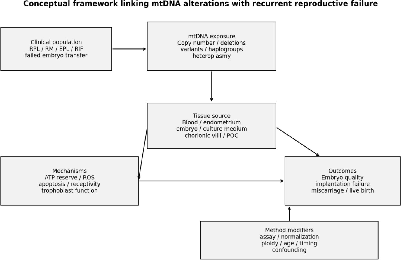
Fig. (2). Tissue-specific conceptual framework linking mtDNA copy-number and genomic-variant pathways with recurrent pregnancy loss, recurrent implantation failure and related reproductive outcomes.
STRENGTHS AND LIMITATIONS
The strengths of this review are that the evidence supporting IVF and direct RPL/RIF evidence was separated, considerations of the amount and number of mtDNA molecules and the level of integrity of the mtDNA sequence were considered separately, the evidence was originally synthesized across the maternal, pregnancy, endometrial, and embryonic compartments of the tissue, and design appropriate JBI or ROBINS-I appraisal was provided while separately considering clinical directness with respect to internal validity Supplementary Table S2A and S2B.
Important limitations remain. Direct RPL/RIF evidence is often few, and are population- and centre-specific, with many positive results being non-replicated and unfunctionalized. The review was not proprioceptively registered, and screening, data extraction and risk-of-bias appraisal were not duplicated separately, so that there is a risk of selection, extraction and appraisal error. Since there is no registration of all prospective protocols, deviations from the original review plan cannot be assessed independently. In the evidence, assay/normalization heterogeneity and inadequate controlling for maternal age and embryo ploidy, and limited external validation, constrict comparability and clinical translation. Other restrictions are the screening and data extraction by a single person, lack of uniformity in mtDNA assays and biological matrices, inadequate replication in direct RPL/RIF studies, and the lack of predictive validity in clinical use.
IMPLICATIONS AND RECOMMENDATIONS
Pregnancy-tissue paired sequencing, ancestry-aware replication in combination with functional annotations of origin from maternal and fatal tissues should be prioritized for future RPL studies. RIF studies need to be reproduced in larger multicentre studies of well-defined endometrial covariates with cycle timing. There should be no universal thresholds for mtDNA in pre-implantation embryos to use until assays are harmonized and incremental predictive value is proven in the cohort of single-euploid embryos they aim to transfer, which is prospectively determined.
Future studies on all the different domains should be explicit on RPL/RIF definition, use validated mtDNA assays, report on the extraction/amplification and normalization procedures, and also control maternal age and embryo ploidy (where applicable) and must include independent validation cohorts with clinically meaningful endpoints preferably live birth.
CONCLUSION
Biologically plausible, but varying associations between mitochondrial genomic changes and reproductive outcomes have been observed. The only consistently reported direct RPL signals are sequence variants, which lack adequate replication or functional validation in populations other than that in which they are found. One small case-control study indicates an association between increased copy numbers of mtDNA and RIF per unit endometrial area, but there is no established limit for diagnosis. Embryo mtDNA content is inconsistent in association on each platform, among each developmental stage and among each euploid-transfer cohort and therefore should not be used as sole criterion when selecting embryos. Thus, biological and epidemiological associations continue to be independent of predictive performance and clinical utility, which are lacking in RPL/RIF. There are a few direct RPL studies that show the best association signal at the sequence level with mtDNA alterations, but the available evidence is still not enough to confirm a diagnostic biomarker. Future prospective large-scale multi-center studies using standardised assays, carefully prespecified confounder control and external validation are needed before clinical implementation.LIST OF ABBREVIATIONS
| ATP | = | Adenosine triphosphate |
| CI | = | Confidence Interval |
| CN | = | Copy Number |
| CNV | = | Copy Number Variant/Variation |
| ddPCR | = | Droplet Digital Polymerase Chain Reaction |
| DHPLC | = | Denaturing high-performance liquid chromatography |
| D-loop | = | Displacement loop (mitochondrial DNA control region) |
| DR | = | Deletion Rate |
| EPL | = | Early Pregnancy Loss |
| ESHRE | = | European Society of Human Reproduction and Embryology |
| eSET | = | Elective Single Embryo Transfer |
| gDNA | = | Genomic DNA |
| IA | = | Induced Abortion |
| ICSI | = | Intracytoplasmic Sperm Injection |
| IRM | = | Idiopathic Recurrent Miscarriage |
| IVF | = | In vitro Fertilisation |
| JBI | = | Joanna Briggs Institute |
| MeSH | = | Medical Subject Headings |
| mtDNA | = | Mitochondrial DNA |
| MT-CO2 | = | Mitochondrially encoded cytochrome c oxidase II |
| MT-ND1 | = | Mitochondrially encoded NADH dehydrogenase 1 |
| mt-tRNA | = | Mitochondrial transfer RNA |
| nDNA | = | Nuclear DNA |
| ND1 / ND2 | = | NADH Dehydrogenase Subunit 1 / 2 (mitochondrially encoded) |
| NGS | = | Next-Generation Sequencing |
| OR | = | Odds Ratio |
| PCR | = | Polymerase Chain Reaction |
| PCR-SSCP | = | Polymerase Chain Reaction–Single-Strand Conformation Polymorphism |
| PEO | = | Population–Exposure–Outcome |
| PGT-A | = | Preimplantation Genetic Testing for Aneuploidy |
| PRISMA | = | Preferred Reporting Items for Systematic Reviews and Meta-Analyses |
| PROSPERO | = | International Prospective Register of Systematic Reviews |
| qPCR | = | Quantitative (real-time) Polymerase Chain Reaction |
| RCOG | = | Royal College of Obstetricians and Gynaecologists |
| RIF | = | Recurrent Implantation Failure |
| RM | = | Recurrent miscarriage |
| ROBINS-I | = | Risk Of Bias in Non-randomised Studies of Interventions |
| ROC | = | Receiver Operating Characteristic |
| ROS | = | Reactive Oxygen Species |
| RPL | = | Recurrent Pregnancy Loss |
| SA | = | Spontaneous Abortion |
| SEFET | = | Single Euploid Frozen Embryo Transfer |
| TE | = | Trophectoderm |
| WGA | = | Whole-Genome Amplification |
| WHO | = | World Health Organization |
AUTHOR’S CONTRIBUTION
I.M. has contributed to the study conceptualization, methodology, data analysis, interpretation of results, and manuscript writing.CONSENT FOR PUBLICATION
Not applicable.AVAILABILITY OF DATA AND MATERIALS
The data will be made available on reasonable request by contacting the corresponding author [I.M.].FUNDING
None.CONFLICT OF INTEREST
The author declares that there is no conflict of interest regarding the publication of this article.ACKNOWLEDGEMENTS
Declared none.DECLARATION OF AI
The author utilized ChatGPT for linguistic refinement and improvement of manuscript readability. All suggested changes were reviewed and verified by the author, who remains fully accountable for the final published content.SUPPLEMENTARY MATERIAL
PRISMA checklist is available as supplementary material on the publisher’s website along with the published article.APPENDIX
Appendix A. Database Search Strategy, Outcomes and Screenshots
The exact database-specific search strings and retrieval counts used for the PRISMA flow diagram are presented below. Screenshots of the database search outcomes are provided in Appendix Figs. A1–A3.
Appendix Table A1. Database search strings and retrieval outcomes by source.
| Database | Exact search string used | Limits/field | Outcome |
| PubMed/MEDLINE | ((“Mitochondrial DNA”[Mesh] OR “mitochondrial DNA”[Title/Abstract] OR mtDNA[Title/Abstract]) AND (“copy number”[Title/Abstract] OR content[Title/Abstract] OR quantification[Title/Abstract] OR variant*[Title/Abstract] OR mutation*[Title/Abstract] OR deletion*[Title/Abstract] OR heteroplasmy[Title/Abstract] OR haplogroup*[Title/Abstract] OR “D-loop”[Title/Abstract] OR “tRNA”[Title/Abstract]) AND (“recurrent pregnancy loss”[Title/Abstract] OR “recurrent miscarriage”[Title/Abstract] OR “repeated pregnancy loss”[Title/Abstract] OR “early pregnancy loss”[Title/Abstract] OR miscarriage[Title/Abstract] OR “spontaneous abortion”[Title/Abstract] OR “recurrent implantation failure”[Title/Abstract] OR “implantation failure”[Title/Abstract] OR “implantation potential”[Title/Abstract] OR implantation[Title/Abstract] OR “embryo transfer”[Title/Abstract] OR “blastocyst transfer”[Title/Abstract] OR “pregnancy outcome”[Title/Abstract] OR “live birth”[Title/Abstract])) | MeSH + Title/Abstract; 2000-2026; English; humans/journal articles during screening. | 164 records |
| Scopus | TITLE-ABS-KEY ( ( “mitochondrial DNA” OR mtDNA ) AND ( “copy number” OR content OR quantification OR variant* OR mutation* OR deletion* OR heteroplasmy OR haplogroup* OR “D-loop” OR “tRNA” ) AND ( “recurrent pregnancy loss” OR “recurrent miscarriage” OR “repeated pregnancy loss” OR “early pregnancy loss” OR miscarriage OR “spontaneous abortion” OR “recurrent implantation failure” OR “implantation failure” OR “implantation potential” OR “embryo transfer” OR “blastocyst transfer” OR “pregnancy outcome” OR “live birth” ) ) AND PUBYEAR > 1999 AND PUBYEAR < 2027 | TITLE-ABS-KEY; PUBYEAR > 1999 and < 2027; English articles. | 333 records |
| Web of Science Core Collection | TS=((“mitochondrial DNA” OR mtDNA) AND (“copy number” OR “mtDNA copy number” OR variant* OR mutation* OR heteroplasmy OR haplogroup* OR deletion*) AND (“recurrent pregnancy loss” OR “recurrent miscarriage” OR “pregnancy loss” OR miscarriage OR “early pregnancy loss” OR “recurrent implantation failure” OR “implantation failure” OR “failed implantation”) AND (human* OR women OR embryo* OR endometrium OR endometrial OR blastocyst* OR trophectoderm)) NOT TS=(review OR “systematic review” OR “meta-analysis”) | Topic/Smart Search; review/meta-analysis terms excluded; article records screened. | 131 records |
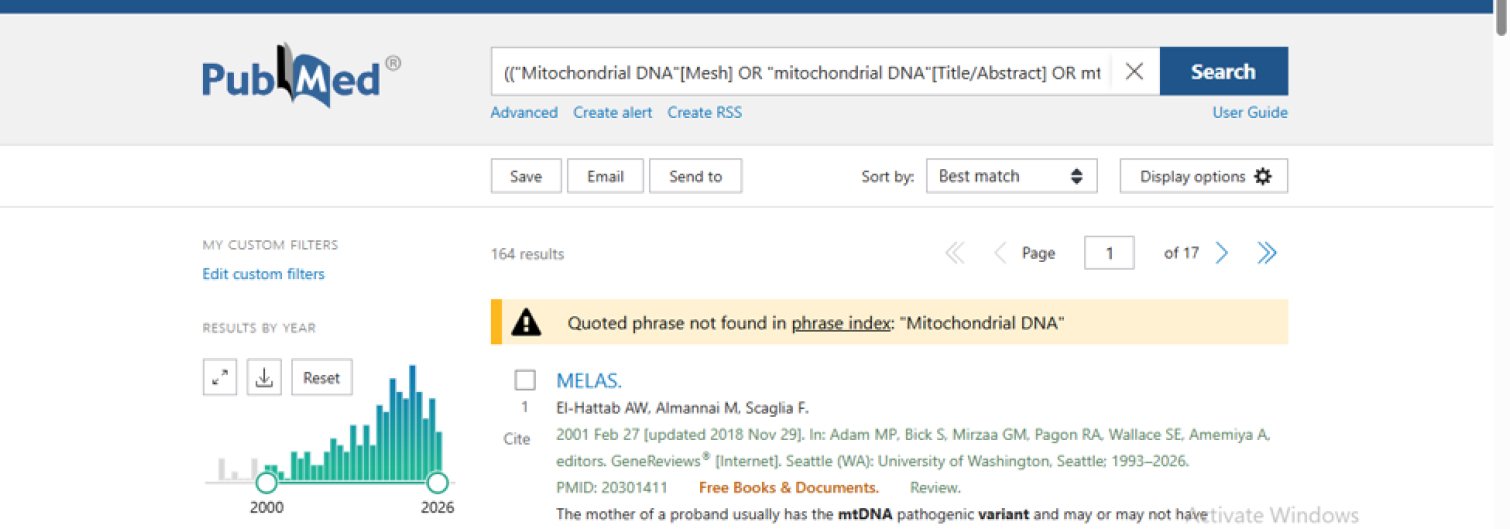
Appendix Fig. (A1). PubMed/MEDLINE search outcome showing 164 records.

Appendix Fig. (A2). Scopus search outcome showing 333 records.

Appendix Fig. (A3). Web of Science Core Collection search outcome showing 131 records.
Appendix B. PRISMA Stepwise Justification and Exclusion Allocation
Appendix B reports the database-wise and merged PRISMA accounting used in the main body. The search identified 628 database records in total (PubMed 164; Scopus 333; Web of Science 131). After 50 duplicates were removed, 578 unique records entered screening. No separate register source contributed records to the final accounting. Screening and full-text exclusions are reported as distinct stages below.
Appendix Table B1. Exclusion reasons at screening and eligibility stages.
| Stage | Exclusion Reason | Number Excluded | Explanation |
| Title/abstract screening | Wrong mitochondrial/reproductive focus | 210 | The record mentioned mitochondria/mtDNA or reproduction but did not address mtDNA copy number, variants or related mitochondrial genomic markers in relevant reproductive failure. |
| Title/abstract screening | Non-primary, non-human or wrong publication type | 125 | Reviews, editorials, letters, conference-only items, animal-only studies or non-human laboratory studies were excluded. |
| Title/abstract screening | No eligible RPL/RIF, pregnancy-loss, implantation, miscarriage or live-birth outcome | 150 | Records did not include a relevant outcome such as RPL, RM, EPL, RIF, implantation failure, embryo-transfer outcome, miscarriage or live birth. |
| Full-text eligibility | No extractable mtDNA marker data | 20 | Full text did not provide extractable mtDNA copy-number, deletion, variant, haplogroup or heteroplasmy data. |
| Full-text eligibility | mtDNA measure not linked to eligible outcome | 23 | The mtDNA measure was not analysed against an eligible clinical or embryological reproductive outcome after full-text assessment. |
| Full-text eligibility | Overlapping cohort, duplicate dataset or insufficient primary outcome data | 15 | Reports used duplicate/overlapping cohorts or lacked sufficient primary outcome data for synthesis. |
| Full-text eligibility | Non-primary publication identified during verification | 1 | Viotti et al. (2017) was a review article and was removed from the primary evidence set. |
Appendix Table B2. Database-wise identification and merged duplicate-removal accounting.
| Source | Records Identified | Duplicate Removal After Merge | Records Entering Screening |
| PubMed/MEDLINE | 164 | – | – |
| Scopus | 333 | – | – |
| Web of Science Core Collection | 131 | – | – |
| Merged total | 628 | 50 | 578 |
Appendix Table B3. Stepwise PRISMA flow after database merging.
| PRISMA Step | Count | Arithmetic / Transition |
| Records identified from databases | 628 | 164 + 333 + 131 = 628 |
| Duplicates removed before screening | 50 | 628 − 50 = 578 |
| Records screened | 578 | — |
| Records excluded at title/abstract | 485 | 578 − 485 = 93 |
| Reports sought for retrieval | 93 | — |
| Reports not retrieved | 5 | 93 − 5 = 88 |
| Reports assessed for eligibility | 88 | — |
| Reports excluded at full text | 59 | 88 − 59 = 29 |
| Studies included in review | 29 | Final included set |
Appendix Table B4. Allocation of the 29 Included Studies by Synthesis Domain
Study-level mapping of included studies according to primary evidence category.
| No. | Study | Year | Country | Primary Population / Source | mtDNA Marker | Assigned Evidence Category |
| 1 | Kaare et al. [28] | 2009 | Finland | Women with recurrent miscarriage + controls | Heteroplasmic mtDNA variants | Direct RPL/RM evidence |
| 2 | Seyedhassani et al. (2010a) [29] | 2010 | Iran | Repeated pregnancy loss + controls | mtDNA deletions and D-loop variants | Direct RPL/RM evidence |
| 3 | Seyedhassani et al. (2010b) [30] | 2010 | Iran | Idiopathic recurrent pregnancy loss mutation screening without controls | mt-tRNA variants | Direct RPL/RM evidence |
| 4 | Vanniarajan et al. [31] | 2011 | India | Women with RPL, fetal tissues, confirmation cohort | Complete mtDNA sequencing | Direct RPL/RM evidence |
| 5 | Colagar et al. [32] | 2013 | Iran | RPL women + controls | ND1 T4216C mutation | Direct RPL/RM evidence |
| 6 | Azadi et al. [33] | 2018 | Iran | RPL women + controls | Complex-I mtDNA variants | Direct RPL/RM evidence |
| 7 | Ye et al. [34] | 2020 | China | Early pregnancy loss chorionic villi + controls | mtDNA copy number and deletion burden | Direct pregnancy-loss evidence |
| 8 | Ji et al. [35] | 2020 | China | Spontaneous abortion vs induced abortion tissues | Copy number and pathogenic variants | Pregnancy-loss tissue evidence |
| 9 | Eker et al. [36] | 2021 | Turkey | RIF women + fertile controls | Endometrial mtDNA copy number | Direct RIF evidence |
| 10 | Mojodi et al. [37] | 2023 | Iran | Idiopathic recurrent miscarriage + controls | mt-tRNA variants | Direct RPL/RM evidence |
Direct / Mixed RIF-RPL Evidence.
| No. | Study | Year | Country | Primary Population / Source | mtDNA Marker | Assigned Evidence Category |
| 11 | Zhou et al. [27] | 2021 | China | Euploid blastocysts including RIF/RM indications | Trophectoderm mtDNA content | Mixed RIF/RM embryo evidence |
| 12 | Łukaszuk et al. [43] | 2022 | Poland | PGT-A embryos including RIF/RM indications | Trophectoderm mtDNA/nDNA ratio | Mixed RIF/RM embryo evidence |
| 13 | Brenner et al. [48] | 2000 | USA | RIF patients after ooplasmic transfer | Donor-recipient mtDNA heteroplasmy | RIF intervention evidence |
| 14 | Barritt et al. [49] | 2001 | USA | Offspring after ooplasmic transplantation | Donor/recipient mtDNA inheritance | RIF intervention evidence |
Contextual IVF / Embryo Transfer / Implantation / Live Birth Evidence.
| No. | Study | Year | Country | Primary Population / Source | mtDNA Marker | Assigned Evidence Category |
| 15 | Fragouli et al. [22] | 2017 | UK/USA | Transferred euploid blastocysts | Trophectoderm mtDNA | Contextual IVF evidence |
| 16 | Ravichandran et al. [23] | 2017 | USA | Euploid blastocysts from IVF clinics | Trophectoderm mtDNA | Contextual IVF evidence |
| 17 | Treff et al. [24] | 2017 | USA | Sibling euploid blastocyst comparison | Trophectoderm mtDNA | Contextual IVF evidence |
| 18 | Victor et al. [25] | 2017 | USA/UK | Blastocysts after PGT-A | Adjusted mtDNA quantification | Contextual IVF evidence |
| 19 | Lee et al. [26] | 2019 | Taiwan | Infertile couples and transferred blastocysts | Adjusted embryo mtDNA quantity | Contextual IVF evidence |
| 20 | Lledó et al. [38] | 2018 | Spain | Euploid single embryo transfers | Blastocyst mtDNA analysis | Contextual IVF evidence |
| 21 | Diez-Juan et al. [39] | 2015 | Spain | Euploid embryo transfers | Embryo mtDNA content | Contextual IVF evidence |
| 22 | Fragouli et al. [40] | 2015 | UK/USA | Embryos undergoing genetic testing | Embryo/trophectoderm mtDNA | Contextual IVF evidence |
| 23 | Stigliani et al. [41] | 2014 | Italy | Spent culture medium samples | Extracellular mtDNA/gDNA ratio | Contextual IVF evidence |
| 24 | Klimczak et al. [42] | 2018 | USA | Blastocyst biopsy dataset | Embryonic mtDNA content | Contextual IVF evidence |
| 25 | Podolak et al. [44] | 2022 | Poland | Cleavage-stage embryos | Embryo mtDNA copy number | Contextual embryo evidence |
| 26 | El-Damen et al. [45] | 2021 | UAE | Single euploid frozen embryo transfers | Blastocyst mtDNA content | Contextual IVF evidence |
| 27 | Wang et al. [46] | 2021 | China | Euploid blastocysts | Trophectoderm mtDNA content | Contextual IVF evidence |
| 28 | Chuang et al. [47] | 2023 | Taiwan | Euploid single embryo transfers | Embryo/trophectoderm mtDNA content | Contextual IVF evidence |
| 29 | Victor et al. [50] | 2019 | USA/UK | Transferred blastocysts with live birth outcomes | Trophectoderm mtDNA content | Contextual IVF evidence |
REFERENCES
[1] World Health Organization. Infertility prevalence estimates, 1990–2021. Geneva: World Health Organization; 2023. Available from: https://www.who.int/publications/i/item/9789240068315
[2] Cox CM, Thoma ME, Tchangalova N, Mburu G, Bornstein MJ, Johnson CL, Kiarie J. Infertility prevalence and the methods of estimation from 1990 to 2021: a systematic review and meta-analysis. Hum Reprod Open. 2022;2022(4): hoac051.
https://doi.org/10.1093/hropen/hoac051
[3] Quenby S, Gallos ID, Dhillon-Smith RK, Podesek M, Stephenson MD, Fisher J, et al. Miscarriage matters: the epidemiological, physical, psychological, and economic costs of early pregnancy loss. Lancet. 2021;397(10285):1658–1667.
https://doi.org/10.1016/S0140-6736(21)00682-6
[4] ESHRE Guideline Group on RPL, Bender Atik R, Christiansen OB, Elson J, Kolte AM, Lewis S, et al. ESHRE guideline: recurrent pregnancy loss: an update in 2022. Hum Reprod Open. 2023;2023(1).
https://doi.org/10.1093/hropen/hoad002
[5] Regan L, Rai R, Saravelos S, Li TC. Recurrent miscarriage: Green-top Guideline No. 17. BJOG. 2023;130(12)–e39.
https://doi.org/10.1111/1471-0528.17515
[6] ESHRE Working Group on Recurrent Implantation Failure, Cimadomo D, de Los Santos MJ, Griesinger G, Lainas G, Le Clef N, et al. ESHRE good practice recommendations on recurrent implantation failure. Hum Reprod Open. 2023;2023(3).
https://doi.org/10.1093/hropen/hoad023
[7] Practice Committee of the American Society for Reproductive Medicine. Evaluation and treatment of recurrent pregnancy loss: a committee opinion. Fertil Steril. 2012;98(5):1103–1111.
https://doi.org/10.1016/j.fertnstert.2012.06.048
[8] Babayev E, Seli E. Oocyte mitochondrial function and reproduction. Curr Opin Obstet Gynecol. 2015;27(3):175–181.
https://doi.org/10.1097/GCO.0000000000000164
[9] May-Panloup P, Boucret L, Chao de la Barca JM, Desquiret-Dumas V, Ferré-L’Hotellier V, Morinière C, et al. Ovarian ageing: the role of mitochondria in oocytes and follicles. Hum Reprod Update. 2016;22(6):725–743.
https://doi.org/10.1093/humupd/dmw028
[10] St John JC, Okada T, Andreas E, Penn A. The role of mtDNA in oocyte quality and embryo development. Mol Reprod Dev. 2023;90(7):621–633.
https://doi.org/10.1002/mrd.23640
[11] Taylor RW, Turnbull DM. Mitochondrial DNA mutations in human disease. Nat Rev Genet. 2005;6(5):389–402.
https://doi.org/10.1038/nrg1606
[12] Wai T, Ao A, Zhang X, Cyr D, Dufort D, Shoubridge EA. The role of mitochondrial DNA copy number in mammalian fertility. Biol Reprod. 2010;83(1):52–62.
https://doi.org/10.1095/biolreprod.109.080887
[13] Stewart JB, Chinnery PF. The dynamics of mitochondrial DNA heteroplasmy: implications for human health and disease. Nat Rev Genet. 2015;16(9):530–542.
https://doi.org/10.1038/nrg3966
[14] Stewart JB, Chinnery PF. Extreme heterogeneity of human mitochondrial DNA from organelles to populations. Nat Rev Genet. 2021;22(2):106–118.
https://doi.org/10.1038/s41576-020-00284-x
[15] Burton GJ, Jauniaux E. Placental oxidative stress: from miscarriage to preeclampsia. J Soc Gynecol Investig. 2004;11(6):342–352.
https://doi.org/10.1016/j.jsgi.2004.03.003
[16] Duhig K, Chappell LC, Shennan AH. Oxidative stress in pregnancy and reproduction. Obstet Med. 2016;9(3):113–116.
https://doi.org/10.1177/1753495X16648495
[17] Zejnullahu VA, Zejnullahu VA, Kosumi E. The role of oxidative stress in patients with recurrent pregnancy loss: a review. Reprod Health. 2021;18:207.
https://doi.org/10.1186/s12978-021-01257-x
[18] Munn Z, Stern C, Aromataris E, Lockwood C, Jordan Z. What kind of systematic review should I conduct? A proposed typology and guidance for systematic reviewers in the medical and health sciences. BMC Med Res Methodol. 2018;18(1):5.
https://doi.org/10.1186/s12874-017-0468-4
[19] Page MJ, McKenzie JE, Bossuyt PM, Boutron I, Hoffmann TC, Mulrow CD, et al. The PRISMA 2020 statement: an updated guideline for reporting systematic reviews. BMJ. 2021;372.
https://doi.org/10.1136/bmj.n71
[20] Aromataris E, Munn Z, editors. JBI Manual for Evidence Synthesis. JBI; 2020.
https://doi.org/10.46658/JBIMES-20-01
[21] Sterne JAC, Hernán MA, Reeves BC, Savović J, Berkman ND, Viswanathan M, et al. ROBINS-I: a tool for assessing risk of bias in non-randomised studies of interventions. BMJ. 2016;355.
https://doi.org/10.1136/bmj.i4919
[22] Fragouli E, McCaffrey C, Ravichandran K, Spath K, Grifo JA, Munné S, et al. Clinical implications of mitochondrial DNA quantification on pregnancy outcomes: a blinded prospective non-selection study. Hum Reprod. 2017;32(11):2340–2347.
https://doi.org/10.1093/humrep/dex292
[23] Ravichandran K, McCaffrey C, Grifo J, Morales A, Perloe M, Munné S, Wells D, Fragouli E. Mitochondrial DNA quantification as a tool for embryo viability assessment: retrospective analysis of data from single euploid blastocyst transfers. Hum Reprod. 2017;32(6):1282–1292.
https://doi.org/10.1093/humrep/dex070
[24] Treff NR, Zhan Y, Tao X, Olcha M, Han M, Rajchel J, et al. Levels of trophectoderm mitochondrial DNA do not predict the reproductive potential of sibling embryos. Hum Reprod. 2017;32(4):954–962.
https://doi.org/10.1093/humrep/dex034
[25] Victor AR, Brake AJ, Tyndall JC, Griffin DK, Zouves CG, Barnes FL, Viotti M. Accurate quantitation of mitochondrial DNA reveals uniform levels in human blastocysts irrespective of ploidy, age, or implantation potential. Fertil Steril. 2017;107(1):34–42.e3.
https://doi.org/10.1016/j.fertnstert.2016.09.028
[26] Lee YX, Chen CH, Lin SY, Lin YH, Tzeng CR. Adjusted mitochondrial DNA quantification in human embryos may not be applicable as a biomarker of implantation potential. J Assist Reprod Genet. 2019;36(9):1855–1865.
https://doi.org/10.1007/s10815-019-01542-6
[27] Zhou X, Liu X, Shi W, Ye M, Chen S, Xu C. Mitochondrial DNA content may not be a reliable screening biomarker for live birth after single euploid blastocyst transfer. Front Endocrinol (Lausanne). 2021;12:762976.
https://doi.org/10.3389/fendo.2021.762976
[28] Kaare M, Götz A, Ulander VM, Ariansen S, Kaaja R, Suomalainen A, Aittomäki K. Do mitochondrial mutations cause recurrent miscarriage? Mol Hum Reprod. 2009;15(5):295–300.
https://doi.org/10.1093/molehr/gap021
[29] Seyedhassani SM, Houshmand M, Kalantar SM, Modabber G, Aflatoonian A. No mitochondrial DNA deletions but more D-loop point mutations in repeated pregnancy loss. J Assist Reprod Genet. 2010a;27(11):641–648.
https://doi.org/10.1007/s10815-010-9435-2
[30] Seyedhassani SM, Houshmand M, Kalantar SM, Aflatoonian A, Modabber G, Hadipour F, Fallahzadeh MH. The point mutations of mitochondrial tRNA threonine and proline in idiopathic repeated pregnancy loss. Iran J Reprod Med. 2010b;8(1):45–50. Available from: https://utoronto.scholaris.ca/items/04570e11-00a5-4b37-a093-fb6080176bfa (Accessed on: 13-10-2026).
[31] Vanniarajan A, Govindaraj P, Carlus SJ, Aruna M, Aruna P, Kumar A, et al. Mitochondrial DNA variations associated with recurrent pregnancy loss among Indian women. Mitochondrion. 2011;11(3):450–456.
https://doi.org/10.1016/j.mito.2011.01.002
[32] Colagar AH, Mosaieby E, Seyedhassani SM, Mohajerani M, Arasteh A, Kamalidehghan B, Houshmand M. T4216C mutation in NADH dehydrogenase I gene is associated with recurrent pregnancy loss. Mitochondrial DNA. 2013;24(5):610–612.
https://doi.org/10.3109/19401736.2013.772150
[33] Azadi A, Seo DJ, Jafari Sasansara H, Van Haute M. Mitochondrial DNA variations are associated with recurrent pregnancy loss. Mitochondrial DNA A DNA Mapp Seq Anal. 2018;29(5):674–678.
https://doi.org/10.1080/24701394.2017.1350854
[34] Ye M, Shi W, Hao Y, Zhang L, Chen S, Wang L, He X, Li S, Xu C. Associations of mitochondrial DNA copy number and deletion rate with early pregnancy loss. Mitochondrion. 2020;55:48–53.
https://doi.org/10.1016/j.mito.2020.07.006
[35] Ji L, Liao T, Yang J, Su H, Song J, Qian K. Deep sequencing shows that accumulation of potentially pathogenic mtDNA mutations rather than mtDNA copy numbers may be associated with early embryonic loss. J Assist Reprod Genet. 2020;37(9):2181–2188.
https://doi.org/10.1007/s10815-020-01893-5
[36] Eker C, Basdas R, Karamustafaoglu Balci B, Bastu E, Gunel T. The genomic analysis of endometrial mitochondrial DNA copy number variation on recurrent implantation failure. J Gynecol Obstet Hum Reprod. 2021;50(2):101945.
https://doi.org/10.1016/j.jogoh.2020.101945
[37] Mojodi E, Mehrjardi AM, Naeimzadeh Y, Ghasemi N, Falahati A, Moshtaghioun SM. The sequence variation of mitochondrial tRNA tyrosine and cysteine among Iranian women with idiopathic recurrent miscarriage: a case-control study. Int J Reprod Biomed. 2023;21(7):567–576.
https://doi.org/10.18502/ijrm.v21i7.13894
[38] Lledó B, Ortiz JA, Morales R, García-Hernández E, Ten J, Bernabeu A, Llácer J, Bernabeu R. Comprehensive mitochondrial DNA analysis and IVF outcome. Hum Reprod Open. 2018;2018(4).
https://doi.org/10.1093/hropen/hoy023
[39] Diez-Juan A, Rubio C, Marin C, Martinez S, Al-Asmar N, Riboldi M, Díaz-Gimeno P, Valbuena D, Simón C. Mitochondrial DNA content as a viability score in human euploid embryos: less is better. Fertil Steril. 2015;104(3):534–541.e1.
https://doi.org/10.1016/j.fertnstert.2015.05.022
[40] Fragouli E, Spath K, Alfarawati S, Kaper F, Craig A, Michel CE, Kokocinski F, Cohen J, Munne S, Wells D. Altered levels of mitochondrial DNA are associated with female age, aneuploidy, and provide an independent measure of embryonic implantation potential. PLoS Genet. 2015;11(6).
https://doi.org/10.1371/journal.pgen.1005241
[41] Stigliani S, Persico L, Lagazio C, Anserini P, Venturini PL, Scaruffi P. Mitochondrial DNA in Day 3 embryo culture medium is a novel, non-invasive biomarker of blastocyst potential and implantation outcome. Mol Hum Reprod. 2014;20(12):1238–1246.
https://doi.org/10.1093/molehr/gau086
[42] Klimczak AM, Pacheco LE, Lewis KE, Massahi N, Richards JP, Kearns WG, Saad AF, Crochet JR. Embryonal mitochondrial DNA: relationship to embryo quality and transfer outcomes. J Assist Reprod Genet. 2018;35(5):871–877.
https://doi.org/10.1007/s10815-018-1147-z
[43] Lukaszuk K, Podolak A. Does trophectoderm mitochondrial DNA content affect embryo developmental and implantation potential? Int J Mol Sci. 2022;23(11):5976.
https://doi.org/10.3390/ijms23115976
[44] Podolak A, Liss J, Kiewisz J, Pukszta S, Cybulska C, Rychlowski M, et al. Mitochondrial DNA copy number in cleavage stage human embryos—impact on infertility outcome. Curr Issues Mol Biol. 2022;44(1):273–287.
https://doi.org/10.3390/cimb44010020
[45] El-Damen A, Elkhatib I, Bayram A, Arnanz A, Abdala A, Samir S, Lawrenz B, De Munck N, Fatemi HM. Does blastocyst mitochondrial DNA content affect miscarriage rate in patients undergoing single euploid frozen embryo transfer? J Assist Reprod Genet. 2021;38(3):595–604.
https://doi.org/10.1007/s10815-020-02050-8
[46] Wang J, Diao Z, Zhu L, Zhu J, Lin F, Jiang W, et al. Trophectoderm mitochondrial DNA content associated with embryo quality and Day-5 euploid blastocyst transfer outcomes. DNA Cell Biol. 2021;40(5):643–651.
https://doi.org/10.1089/dna.2020.6271
[47] Chuang TH, Chen CY, Kuan CS, Lai HH, Hsieh CL, Lee MJ, et al. Reduced mitochondrial DNA content correlate with poor clinical outcomes in cryotransfers with day 6 single euploid embryos. Front Endocrinol (Lausanne). 2022;13:1066530.
https://doi.org/10.3389/fendo.2022.1066530
[48] Brenner CA, Barritt JA, Willadsen S, Cohen J. Mitochondrial DNA heteroplasmy after human ooplasmic transplantation. Fertil Steril. 2000;74(3):573–578.
https://doi.org/10.1016/S0015-0282(00)00681-6
[49] Barritt JA, Brenner CA, Malter HE, Cohen J. Mitochondria in human offspring derived from ooplasmic transplantation: brief communication. Hum Reprod. 2001;16(3):513–516.
https://doi.org/10.1093/humrep/16.3.513
[50] Victor AR, Griffin DK, Gardner DK, Brake AJ, Zouves CG, Barnes FL, Viotti M. Births from embryos with highly elevated levels of mitochondrial DNA. Reprod Biomed Online. 2019;39(3):403–412.
https://doi.org/10.1016/j.rbmo.2019.03.214